1800 605-5127
 +61 (0)8 8352 7711
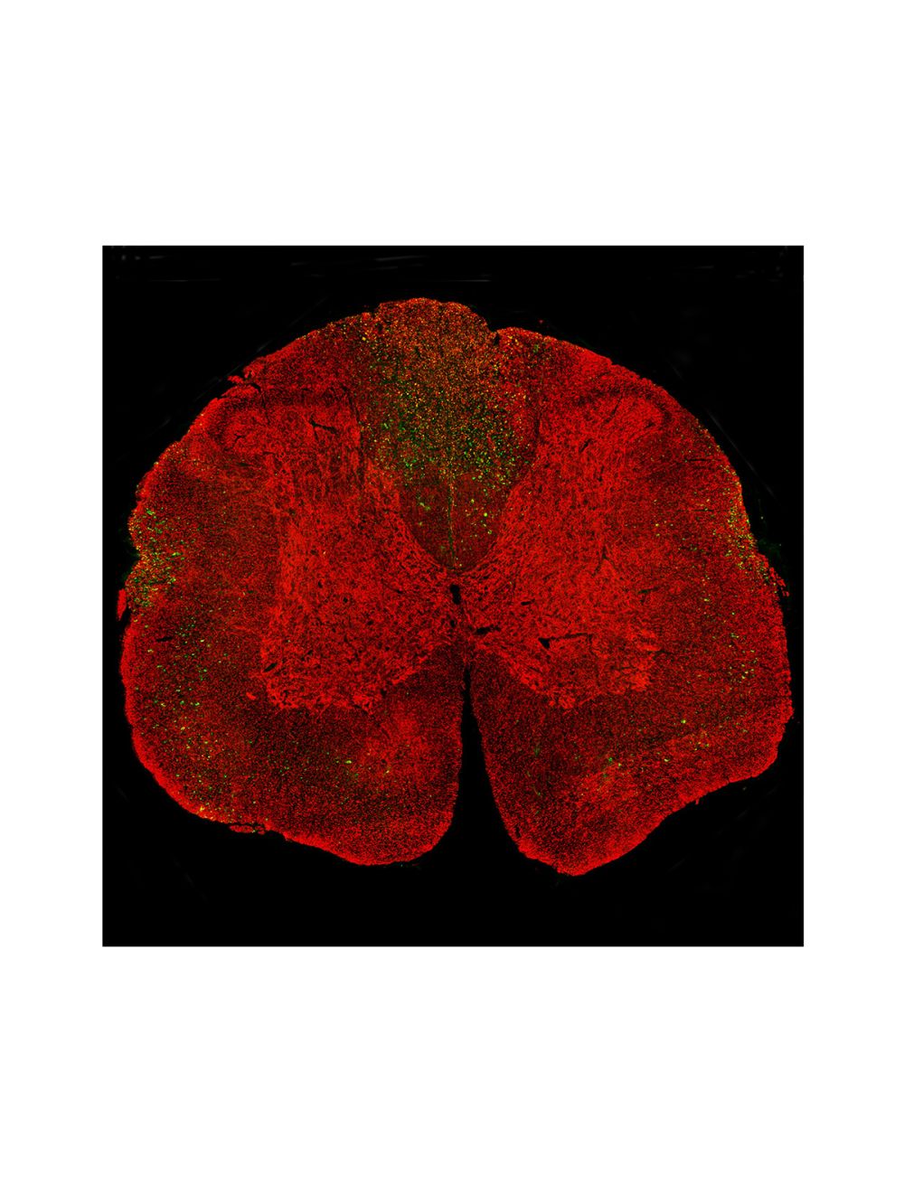

Neurofilament light polypeptide (NF-L), Mouse Monoclonal Antibody (1B11)
- Product Name Neurofilament light polypeptide (NF-L), Mouse Monoclonal Antibody (1B11)
-
Product Description
Mouse anti-Neurofilament light polypeptide (NF-L), Monoclonal Antibody (Unconjugated), Clone 1B11, suitable for WB and Immunostaining.
- Alternative Names NFL, Neurofilament light, NF-L degenerative, NEFL, CMT1F, CMT2E, NF-L, NF68, 1D44, PPP1R110, neurofilament light, CMTDIG, neurofilament light chain
- Application(s) IF, ICC, IHC, WB
- Antibody Host Mouse
- Antibody Type Monoclonal
- Specificity Species cross-reactivity includes human, rat, mouse, cow, and pig.
- Species Reactivity Bovine, Human, Mouse, Pig, Rat
- Immunogen Description The antibody has been made against a preparation of NF-L protein purified from pig spinal cord. This antibody binds to amino acids 316-370 of human NF-L.
- Conjugate Unconjugated
- Purity Description Protein G purified
- Regulatory Status For research use only.
Product Info
-
Product Description
Mouse anti-Neurofilament light polypeptide (NF-L), Monoclonal Antibody (Unconjugated), Clone 1B11, suitable for WB and Immunostaining.
-
Related Products
Neurofilament light polypeptide (NF-L), Chicken Polyclonal Antibody
Neurofilament light polypeptide (NF-L), DG-Sensor™, Chicken Polyclonal Antibody
Neurofilament light polypeptide (NF-L), Mouse Monoclonal Antibody (DA2)
Neurofilament light polypeptide (NF-L), Mouse Monoclonal Antibody (7D1)
Neurofilament light polypeptide (NF-L), Mouse Monoclonal Antibody (6H112)
Neurofilament light polypeptide (NF-L), DG-Sensor™, Mouse Monoclonal Antibody (6H63)
Neurofilament light polypeptide (NF-L), DG-Sensor™, Mouse Monoclonal Antibody (1D44)
Neurofilament light polypeptide (NF-L), Rabbit Polyclonal Antibody
Neurofilament light polypeptide, C-terminus, (NF-L-Ct), Rabbit Polyclonal Antibody
Neurofilament light polypeptide (NF-L), DG-Sensor™, Rabbit Polyclonal Antibody
- Application(s) IF, ICC, IHC, WB
-
Application Details
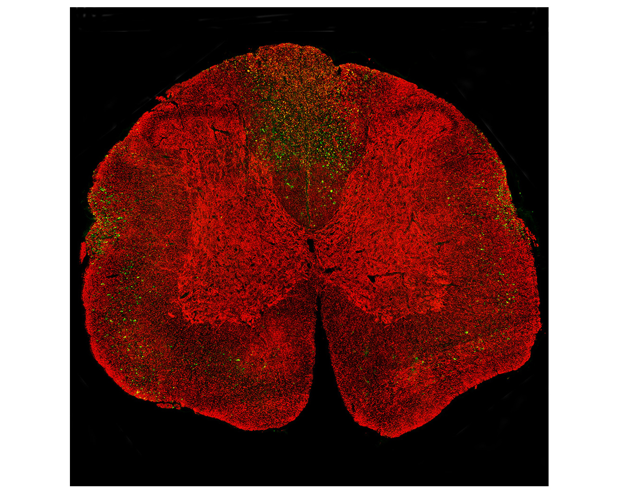
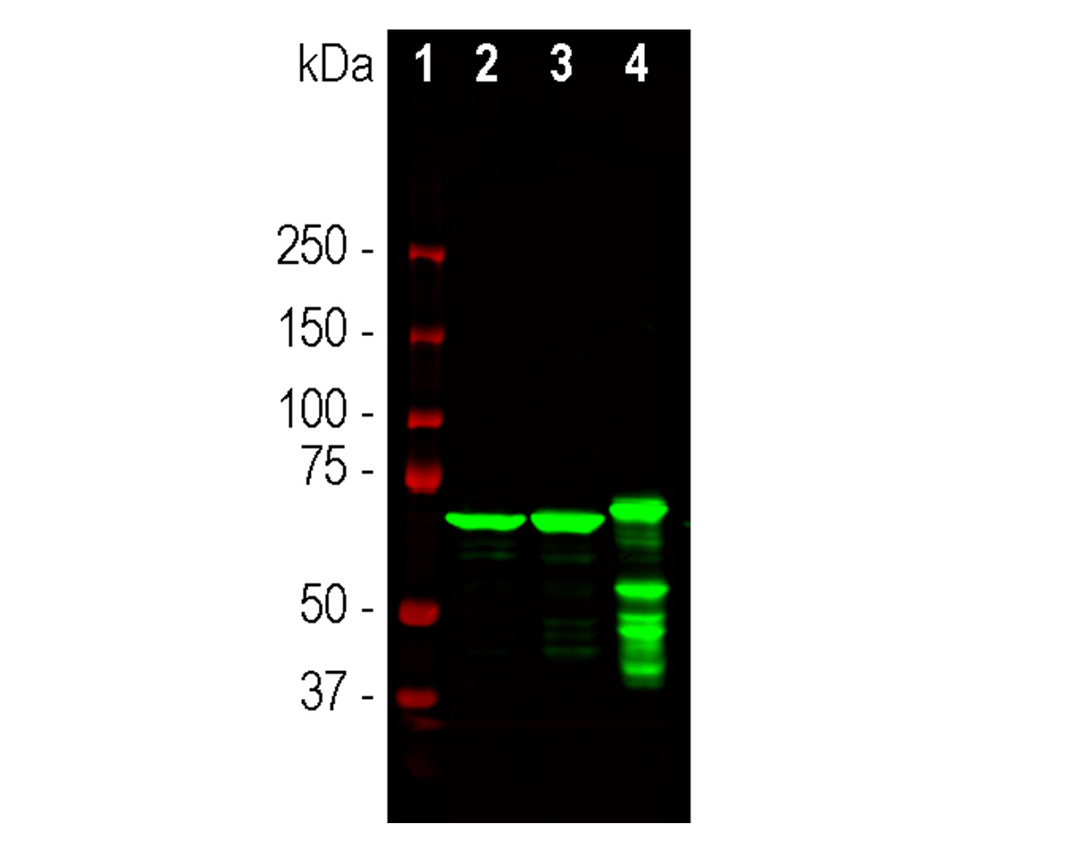
Western blot (WB), Immunocytochemistry (ICC) / Immunofluorescence (IF), Immunohistochemistry (IHC). A dilution of 1:10,000 - 1:20,000 is recommended for WB. A dilution of 1:2,000 is recommended for ICC/IF and IHC. The antibody recognizes NF-L in reduced westerns regardless of the disease state.
Using standard fluorescent antibody methods, a 1: 1000-2,000 dilution is recommended for ICC/IF. For example, block and permeabilize sections in 5-10% normal goat serum or serum of the species the secondary antibodies were made in, in PBS plus 1% Triton-100 (PBST) for 1 hour with slight agitation, followed by primary antibody incubations and fluorescent secondary identification. High primary antibody dilutions require refrigerated, overnight incubations for best results. Recommended fixation is 4% PFA fixed, frozen tissue 20-50 microns; other fixation methods have not been tested and are not recommended at this time.
Degeneration-specific detection is fixation, antigen recovery, and concentration-dependent. The epitope detected a proprietary recombinant immunogen based on the Coil 2 region of human NF-L. This peptide epitope can be uncovered in degenerating cells but not normal cells. However, Clone 1B11 can bind normal neurofilaments when used at high concentrations but shows strong binding to degenerated material at lower antibody concentrations. The specific reactivity of this epitope is sensitive to tissue treatments and could become exposed in healthy cells under some conditions. For example, treatment of the fixed tissue with high temperatures, proteinase, or other denaturants may cause the reactive epitope to become exposed in healthy cells, leading to a false positive. Biosensis recommends experimenting with treated and untreated tissues when first using these antibodies if degeneration specificity is desired. The exact conditions and dilutions must be determined experimentally by the end user.
This antibody will detect NL-L protein in paraffin-embedded rodent tissues; however, the degeneration-specific detection can be problematic in paraffin tissues, particularly if Heat-Induced Epitope Retrieval (HIER), or other common antigen recovery methods are used. This is because the reactive epitope, which is covered in healthy cells but exposed in degenerative cells, could become accessible to the antibody in healthy cells, leading to false positives. For this reason, paraffin-embedded tissues are not recommended if degeneration-specific detection is desired. This antibody was made against NF-L purified from pig spinal cord and binds to amino acids 316-370 of human NF-L
- Target Neurofilament light polypeptide (NF-L)
- Specificity Species cross-reactivity includes human, rat, mouse, cow, and pig.
- Target Host Species Pig
- Species Reactivity Bovine, Human, Mouse, Pig, Rat
- Antibody Host Mouse
- Antibody Type Monoclonal
- Antibody Isotype IgG1
- Clone Name 1B11
- Conjugate Unconjugated
- Immunogen Description The antibody has been made against a preparation of NF-L protein purified from pig spinal cord. This antibody binds to amino acids 316-370 of human NF-L.
- Purity Description Protein G purified
- Format Lyophilized from PBS buffer pH 7.2-7.6 with 0.1% trehalose, and sodium azide
- Reconstitution Instructions Spin vial briefly before opening. Reconstitute with 100 µL sterile-filtered, ultrapure water to achieve a 1 mg/mL concentration. Centrifuge to remove any insoluble material.
- Storage Instructions Store lyophilized antibody at 2-8°C After reconstitution of lyophilized antibody, aliquot and store at -20°C for a higher stability. Avoid freeze-thaw cycles. Store at 4°C for up to one month for short term storage and frequent use.
- Batch Number Please see item label.
- Expiration Date 12 months after date of receipt (unopened vial).
- Alternative Names NFL, Neurofilament light, NF-L degenerative, NEFL, CMT1F, CMT2E, NF-L, NF68, 1D44, PPP1R110, neurofilament light, CMTDIG, neurofilament light chain
- Uniprot Number P07196
- Uniprot Number/Name P07196 (NFL_HUMAN)
- Scientific Background Neurofilaments are composed of three intermediate filament proteins: light (NF-L ~68 kDa), medium (NF-M ~160 kDa) and heavy (NF-H ~200 kDa), found specifically in neurons, which are involved in the maintenance of the neuronal caliber. Neurofilament light (NF68 or NF-L) is the most abundant of the three proteins. (Ref: uniprot.org)
- Shipping Temperature 25°C (ambient)
- UNSPSC CODE 41116161
- Regulatory Status For research use only.