 1800 605-5127
 +61 (0)8 8352 7711
Biosensis Tracing Reagents – Visualize Neurodegenerative Disease Pathways!
Biosensis offers a large variety of established and well-characterized tracing reagents for detecting degenerating neurons (Fluoro-Jade B and C), amyloid plaque (Amylo-Glo and HQ-O), and normal & pathogenic myelin (Black Gold II). These staining reagents are excellent research tools giving maximum flexibility to investigate neurodegenerative disease pathways and disease-modifying drugs.
Biosensis Tracing Reagents exhibit several key advantages:
· Highly published in high impact journals – quality validated by your peers
· Detailed protocols – easy-to-use reagents guaranteed to work
· Excellent technical support – ensuring your experiments will be successful
· Flexibility – compatible with various tissue types, staining techniques and existing antibody immunostaining protocols
Identifying Degenerating Neurons with Fluoro-Jade B and C
The causes and effects of neuronal degeneration are of major interest to a wide variety of neuroscientists. Paralleling this interest is an increasing number of methods applicable to the detection of neuronal degeneration. Fluoro-Jade B (FJB) and C (FJC) tracing reagents stain all degenerating neurons regardless of specific insult or mechanism of cell death.
|
|
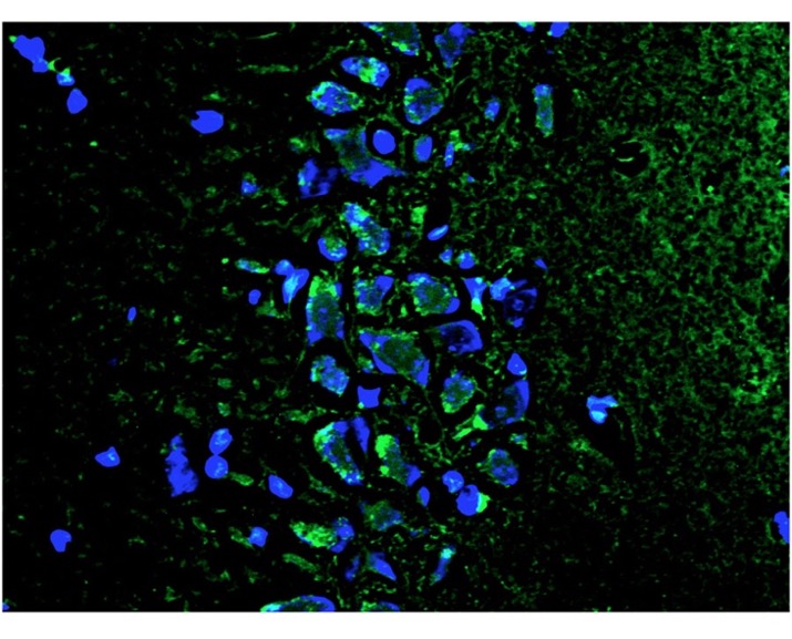
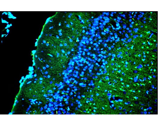
Left: Fluoro-Jade C (FJC) staining (green) of degenerating neurons in the CA3 region of the hippocampus in old diabetic rats. Blue: DAPI nuclear stain. Picture courtesy of Dr. Wang and colleagues (Wang S. et al. (2020). Right: Double-exposure using combined blue and ultraviolet epifluorescence illumination of the superficial layers of the cingulated rat cortex exposed to kainic acid. Layer I contains conspicuous Fluoro-Jade C positive degenerating axon terminals. Layer II contains densely packed DAPI-positive viable granule cells. Layer III contains a mixture of FJC-positive denegerating pyramidal cells and DAPI-positive viable pyramidal cells. Photo is courtesy of Dr. Larry Schmued.
FJC exhibits the greatest signal to background ratio, as well as the highest resolution. This translates to a stain of maximal contrast and affinity for degenerating neurons, and makes it ideal for localising not only degenerating nerve cell bodies, but also distal dendrites, axons and terminals. The dye is highly resistant to fading and is compatible with virtually all histological processing and staining protocols.
The fluorescent dye Fluoro-Jade B (FJB), like its more purified brother FJC, is an anionic fluorescein derivative useful for the histological staining of neurons undergoing degeneration. FJB differs from FJC in that it is a slightly less refined chemical formulation and thus it does not quite provide the same level of signal-to-noise or high resolution as FJC. Nonetheless FJB is still widely used and works very well as a marker of degenerating neurons and even glia (see Damjanac M et al., 2007). FJB operates nearly identically in protocol to that of FJC, and FJB is compatible with several other labelling procedures including immunofluorescent and fluorescent Nissl techniques.
Biosensis Fluoro-Jade B and C Tracing Reagents are available in the following sizes:
TR-100-FJ, Fluoro-Jade C (FJC) Ready-to-Dilute (RTD) Staining Kit (40 mL)
TR-100-FJT, Fluoro-Jade C (FJC) Ready-to-Dilute (RTD) Staining Kit, Trial Size (20 mL)
TR-150-FJ, Fluoro-Jade B (FJB) Powder (30 mg)
TR-160-FJ, Fluoro-Jade C (FJC) Powder (30 mg)
Note: Fluoro-Jade B (FJB) Powder (TR-150-FJ) is equivalent to discontinued product AG310 from Merck-Millipore. Fluoro-Jade C (FJC) Powder (TR-160-FJ) is equivalent to discontinued product AG325 from Merck-Millipore.
Identifying Amyloid Plaque with Amylo-Glo and HQ-O
Amylo-Glo Ready-to-Dilute (RTD) Staining Reagent (TR-300-AG) is designed to stain amyloid plaques in tissue sections. This marker has several advantages over other conventional markers such as Thioflavin S and Congo Red because of its unique chemical and spectral properties (Schmued L et al., 2012). Using Amylo-Glo results in a very bright blue UV excitable stain under physiological conditions that will not bleed through when illuminated with other filters. Its brightness makes it ideal for low magnification quantification studies, while its unique excitation/emission profile and mild staining conditions makes it ideal for combination for multiple immunofluorescent labelling studies. Amylo-Glo RTD is compatible with fresh, frozen, and formalin-fixed immunohistochemistry or immunocytochemistry, and it is particularly good for confocal and multiple labelling because of its high fluorescent intensity, high resistance to photo-bleaching, and unique characteristic of UV channel fluorescence.
|
|
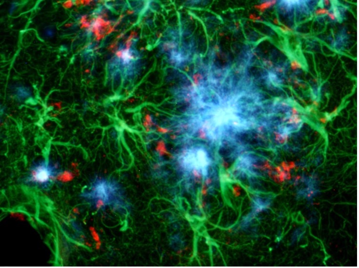
Left: Triple exposure allows for the simultaneous localization of Amylo-Glo positive amyloid plaques (blue), GFAP positive hypertrophied astrocytes (green) and activated microglia (red) in the hippocampus of the AD/Tg mouse. Combined UV, blue and green light illumination. Right: Amylo-Glo labelling of amyloid plaques (blue) with ethidium bromide Nissl (cell body) counterstain (red) in the dentate gyrus region of the hippocampus of the AD/Tg mouse.
The Biosensis Amylo-Glo RTD Amyloid Plaque Stain Reagent with EtBr counter stain (TR-400-AG) kit utilizes an ethidium bromide counter stain for a quick and effective way to visualize cell nuclei and cell bodies of cells, while under UV illumination allowing the assessment of amyloid plaques and cell/tissue positioning in one step.
HQ-O Ready-To-Dilute (RTD) Tracing Reagent is designed to label amyloid plaques in paraffin-embedded or freshly cut frozen tissue sections. As a fluorescent zinc chelator, HQ-O is unique as it takes advantage of the known presence of concentrated zinc in amyloid plaques. Studies with HQ-O revealed that fluorescent plaque-like structures are only seen when synthetic Aβx-42 is aggregated in the presence of zinc. Under blue light excitation, plaque structures appear bright green fluorescent in the brain parenchyma, correlating closely with plaque structures observed following Aβ antibody staining. HQ-O RTD staining reagent is compatible with other fluorophores, such as DAPI, Hoechst and ethidium bromide, as well as fluorescent-labelled antibodies with emission spectra in the blue and/or red emission range of fluorescent microscopes. Due to its zinc-chelating characteristics, HQ-O RTDTM staining reagent may visualize globular structures within blood vessels and intravascular leucocytes.
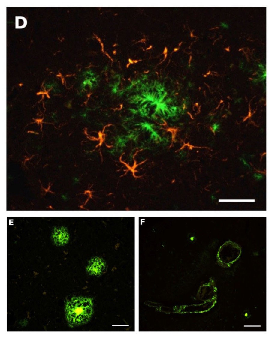
| D: Within the CA4 region of the hippocampus, co-labelling using GFAP antibody demonstrates the relationship between hypertrophied astrocytes (orange) and the amyloid plaques (green) that they typically surround. GFAP-immunoreactivity was visualized with TRITC fluorophore. E: Three amyloid plaques (green) are seen in the cortex of a human with Alzheimer's disease. F: An example of vascular plaques in the cortex of a human AD patient. |
HQ-O RTD staining reagent has multiple advantages over older blue-light exciting stains such as Thioflavin S:
· HQ-O binds to amyloid fibrils and plaques independent of confirmation via unique zinc binding properties
· Exhibits greater contrast and resolution compared to Thioflavin S, Thioflavin T, and Congo Red
· Has tight excitation and emission spectra, with no excitation bleed-through, easily allowing sharp multiple labelling studies
· Uses common blue-light (FITC fluorescence) filters (Emax 475 nm)
· Is compatible with FFPE and frozen tissue sections
· Is EASIER, SHARPER & BRIGHTER than any other non-UV amyloid stain!
Biosensis Amyloid Plaque Tracing Reagents are available in the following sizes:
TR-300-AG, Amylo-Glo RTD Amyloid Plaque Stain Reagent (5 mL)
TR-400-AG, Amylo-Glo RTD Amyloid Plaque Stain Reagent with EtBr counter stain (5 mL)
TR-700-HQO, HQ-O RTD Amyloid Plaque Stain Reagent (40 mL)
Identifying Normal & Pathogenic Myelin with Black-Gold II
Black-Gold II is a haloaurophosphate complex which localises myelin within the central nervous system. The Black Gold II Ready-to-Dilute (RTD) Staining Kit allows to localize myelin, both individual fibres and tracts, along with the option of co-localising cell bodies via the Toluidine Blue counter stain. Black Gold II labelled myelinated fibres appear nearly black while the Toluidine Blue O labelled cellular Nissl bodies are blue under bright field illumination.
Black Gold II can demonstrate and characterise specific myelin changes associated with exposure to diverse neurotoxicants including kainic acid, domoic acid, 3-nitropropionic acid, Fluoro-Gold and isoniazid. Black Gold II can also be combined with other histochemical markers including Nissl stains, retrogradely transported fluorescent tracers and fluorescent markers of neuronal degeneration. The advantages associated with the Black-Gold II include high resolution, high contrast, short histochemical processing time, versatility, and consistent reproducibility.
|
|
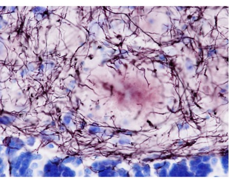

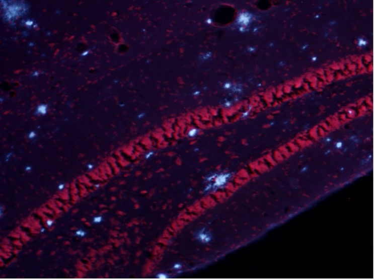
Left: Bright field illumination (60X) of 8-month-old Ad-Tg mouse hippocampus. The myelin pathology can be observed in and around amyloid plaques. The cell bodies of adjacent granule and polymorph cells appear blue, while individual myeliated fibres appear nearly black. Photo is courtesy of Dr. L. Schmued. Right: Human brain Locus K calcitonin gene-related peptide-like immunoreactivity (black) and myelin staining (red, Black-Gold II staining kit, Biosensis) in the medulla oblongata, showing the similar neurochemical and structural organization of the dorsal column nuclei Locus K and the spinal trigeminal substantia gelatinosa (Del Fiacco M. et al., 2018).
Biosensis Normal & Pathogenic Myelin Tracing Reagent is available in the following size:
TR-100-BG, Black-Gold II Myelin Ready-to-Dilute Staining Kit with Toluidine Blue O Counter Stain for identifying Normal & Pathogenic Myelin (10 mL)